
Un jury d’experts
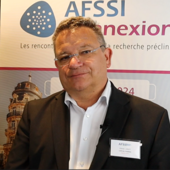
Oliver LOGET
Président & CEO
CapEval Pharma

Joffrey Ducroq
Principal Research Scientist
Charles River Laboratories
Découvrez les sociétés soumettant un poster scientifique pour l’édition 2026
Félicitation à notre lauréat 2026 !
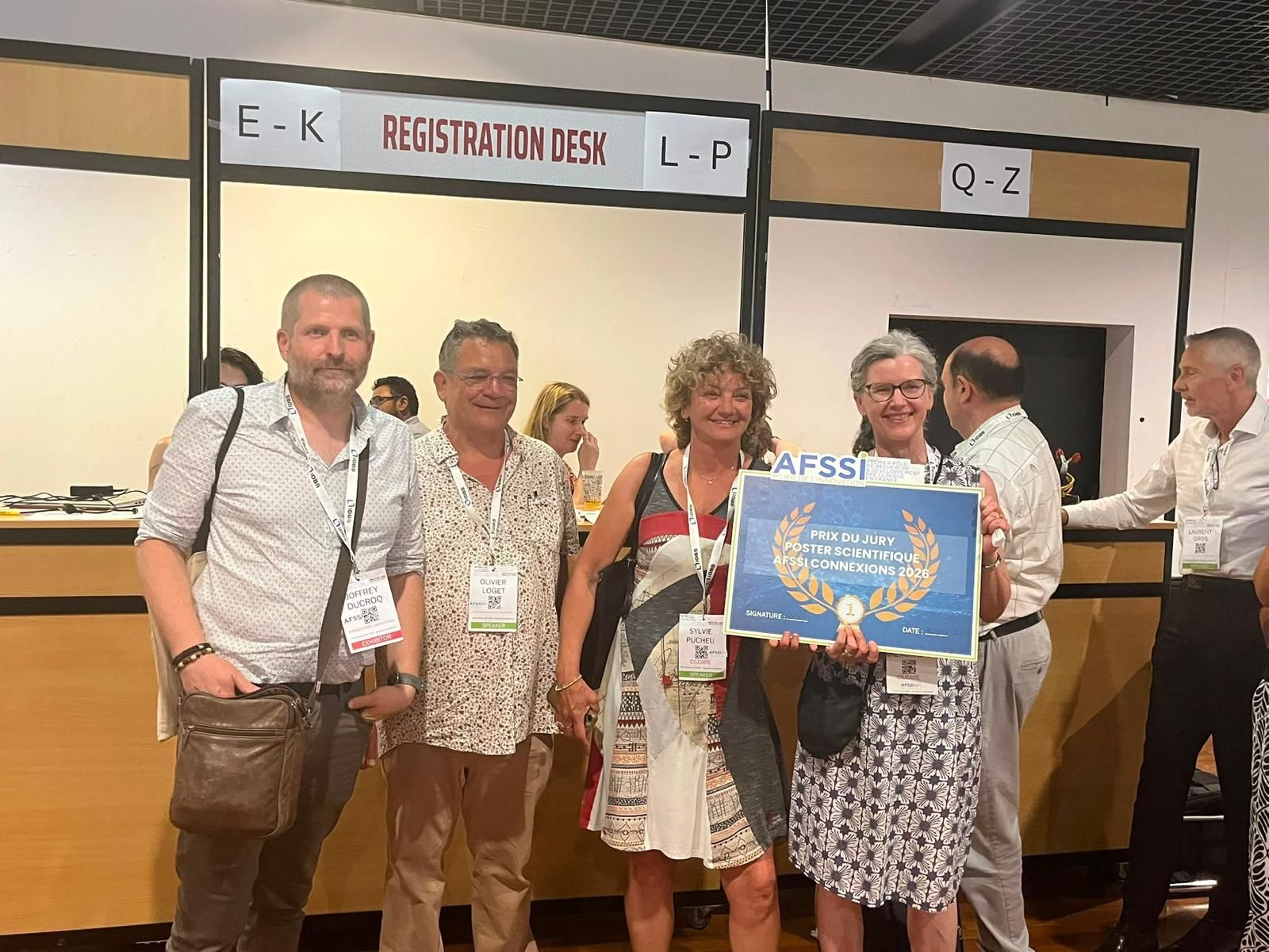
Le prix du jury
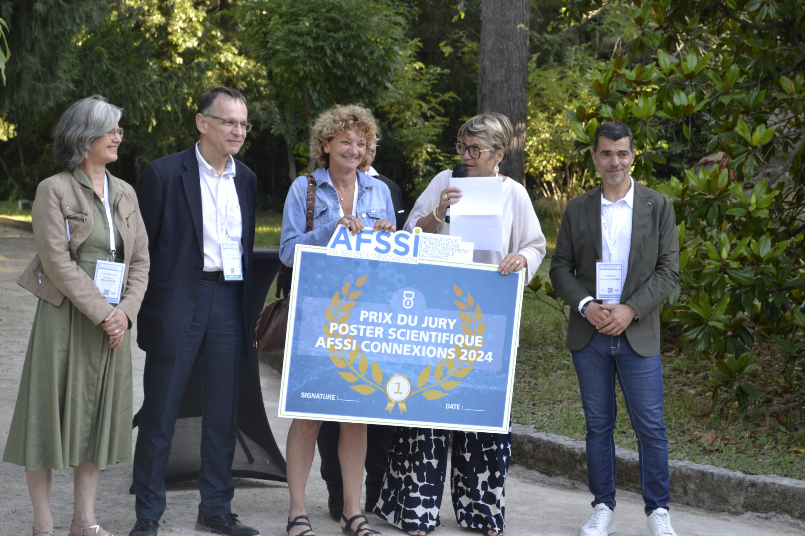
Le jury a décerné le prix du meilleur poster à Cilcare pour son poster intitulé « Innovative collaborative study involving in vitro maturation of oocytes and whole embryo transfer in the mouse », représenté par Susanna Malmström et Sylvie Pucheu.
(Re)découvrez les lauréats 2025
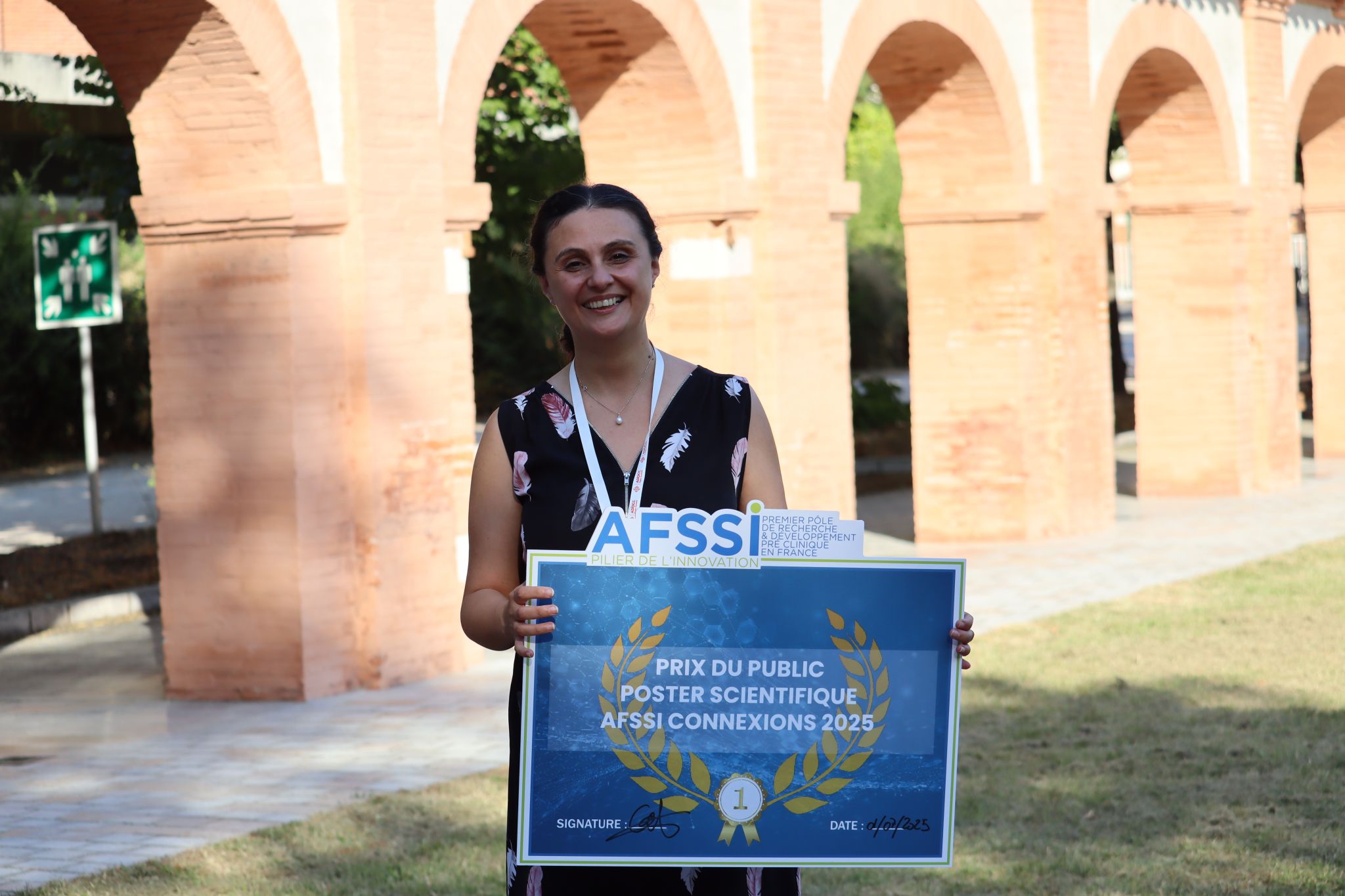
Le prix du public
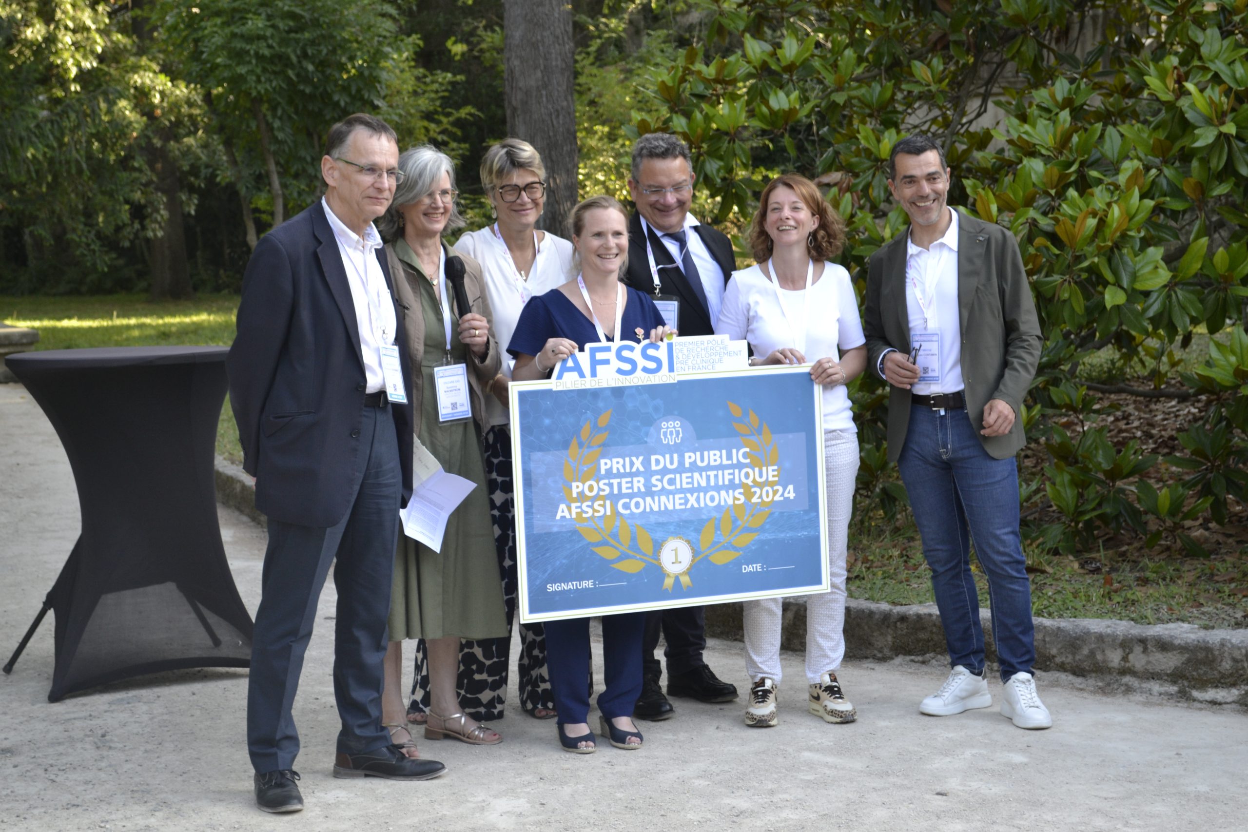
Le public a récompensé G.CLIPS biotech, représenté par Rosie Dawaliby, PhD et Souad GUESSAS pour son poster « Development of conformational and tissue specific antibody against a GPCR as potential drug candidate in immuno-oncology »
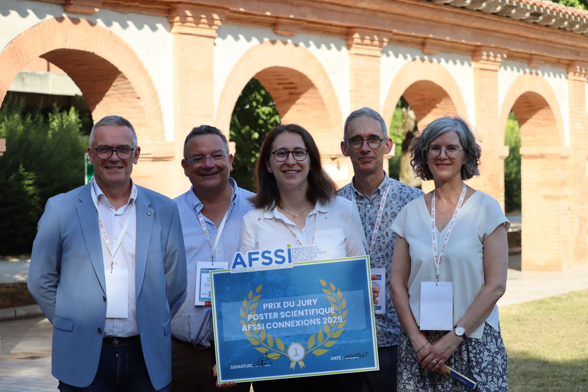
Le prix du jury
Le jury a décerné le prix du meilleur poster à Charles River Laboratories pour son poster intitulé « Innovative collaborative study involving in vitro maturation of oocytes and whole embryo transfer in the mouse », représenté par Marie Bouressam.
CILcare, lauréat du prix du jury
Sensorineural hearing loss as a complication of type-2 Diabetes mellitus: evidence of several cellular and neural impairments.
Abstract
Carolanne COYAT1*, Karine TOUPET1*, Gaëlle NAERT1*, Sylvie PUCHEU1* and Mathieu SCHUE1*.
- Cilcare SAS, 34080 Montpellier France
While retinopathy, nephropathy, and peripheral neuropathy are well-established complications of type 2 diabetes, sensorineural hearing loss is increasingly recognized as a comorbidity of this metabolic disease. Several causes have been suggested, including auditory peripheral neuropathy (synaptopathy), which can lead to early-onset hearing loss. No extensive research has been conducted on preclinical models of type 2 diabetes. Our study aims to better characterize the development of SNHL concomitantly with diabetes biomarkers in a diabetic mouse model.
Genetically modified mice with mutations in the leptin receptor gene (BKS(D)-Leprdb/JOrlRj)2 were monitored from 5 to 13 weeks of age to assess the parameters of their diabetes (blood glucose, glycosylated hemoglobin, biochemical analyses) and their hearing, using auditory evoked potentials and terminal histological analyses of the cochlea.
In this model, Leprdb/Leprdb mice exhibited a phenotype of obesity and hyperglycemia, with fasting blood glucose levels of ~4 g/L compared to 2 g/L in heterozygous control mice. Diabetic mice displayed early-onset hearing loss characterized by a significant increase of auditory brainstem response (ABR) thresholds and a significant decrease of distortion product otoacoustic emission (DPOAE) amplitudes compared to the Leprdb/+ control mice. These data correlated with morphological and histological changes in the cochlea.
Here, we present data from a murine model illustrating the detrimental consequences of type 2 diabetes on hearing. This translational preclinical model may be useful for evaluating the efficacy of drug candidates for the preservation and restoration of hearing in type 2 diabetic patients.
Atlantic Bone Screen et Enterosys, lauréats du prix du public
Décrypter l’Axe Intestin-Articulation : Vers de Nouvelles Stratégies Thérapeutiques ou Préventive pour l’Obésité
Abstract
Dans le contexte de la prévalence croissante de l’obésité et de ses comorbidités, la compréhension des interactions entre l’intestin et les articulations devient cruciale. Enterosys, spécialiste de la communication entre l’intestin et les organes périphériques, en collaboration avec Atlantic Bone Screen, expert en recherche préclinique dans les pathologies osseuses et articulaires, propose une offre de service intégrée pour étudier l’axe intestin-articulation. Cette approche innovante vise à explorer le potentiel thérapeutique de nouveaux actifs dans le traitement des pathologies ostéo-articulaires associées à l’obésité.
Nous avons utilisé un modèle murin, des souris C57BL6J mâles de 9 semaines, soumises à un régime hyperlipidique à 60% pour induire l’obésité. L’étude s’est focalisée sur l’évaluation de divers paramètres : métaboliques (gain de poids, test oral de tolérance au glucose, profils lipidiques plasmatiques, poids des tissus adipeux), intestinaux (marqueurs de la fonction de barrière et perméabilité, analyses histologiques) et articulaires (analyses morphologiques par microtomographie (µCT) et histopathologiques).
Les résultats montrent des modifications significatives des paramètres métaboliques et des altérations des marqueurs de la fonction de barrière intestinale. Des signes d’inflammation et d’atteintes au niveau ostéoarticulaire peuvent être aussi observés, soulignant l’impact de l’obésité sur la santé ostéo-articulaire.
Cette étude met en évidence le potentiel de l’axe intestin-articulation comme cible thérapeutique dans les pathologies métaboliques et dégénératives. Notre offre de service conjointe permettra d’offrir une solution complète pour l’évaluation de l’efficacité d’actifs dans un cadre préclinique, promouvant une approche holistique dans la gestion de l’obésité et ses comorbidités articulaires.
ICI UN VISUEL
Sponsor GOLD
- All
- 1| Stand 16m²
- 2| Visibilité on-line
- 3| Visibilité sur place
- 4| Prise de parole
- 5| Vos accès gold
- 7| Divers
- Sponsor Gold
Accès à une session d’elevator pitch le 1 ou 2 juillet. Présentation express de 2 minutes.
Discours introductif de présentation de votre société (en qualité de mécène) de quelques minutes à l’occasion de la soirée de gala.
- Stand modulaire de 16m²
(Base rectangulaire) - Mise à disposition 2 jours
- 1 table 4 chaises
- 3 chauffeuses, 1 table-basse
- Une banque d’accueil et un tabouret
- Présentoir
- Electricité, accès wifi, éclairage
- Signalétique lettrée
- 1 ou 2 angles ouverts
- 6 pass 2 jours offerts pour vous ou votre réseau (hors cabinets conseils extérieurs)
- Accès 6 personnes à la journée congrès
- Accès 6 personnes à la journée convention d’affaires
- Accès 6 personnes à l’outils de prise de rendez-vous
- Accès 6 personnes à la soirée de gala
- Des invitations supplémentaires à destination de donneurs d’ordre peuvent vous être mises à disposition sur demande
- 2 pages d’insertion publicitaire dans le programme des conférences remis sur place
- Programme (pdf) diffusé en amont au téléchargement sur le site internet et les réseaux sociaux
- Fichier fourni par vos soins
Engagez-vous sur deux éditions et bénéficiez de 30% de remise sur votre formule de sponsoring 2026 !
Chaque partenaire est unique !
Nos équipes seront à l’écoute de vos objectifs et contraintes pour vous proposer une formule adaptée et personnalisée. Votre réussite sera notre réussite !
- Un kakémono disposé sur le site de la manifestation
- Mise à disposition de vos plaquettes en libre service
- L’animation d’une session de présentation
Sous réserve d’acceptation par l’organisateur - Mention de votre intervention sur le programme de la manifestation
- Une chambre pour deux personnes dans un hôtel 3 étoiles
- Votre logo, film ou PowerPoint diffusé sur un écran plasma lors de l’événement
- Votre logo affiché sur uneide « sponsor » à l’occasion du congrès et des elevator pitches
- Annonce de notre sponsoring sur Twitter et LinkedIn
- Relais par nos partenaires
- Parution d’un post contextuel relatif à votre expertise sectorielle (en lien avec les thèmes AFSSI Connexions)
- Transmission d’un visuel personnalisé pour illustrer la diffusion de votre participation sur les réseaux sociaux
- Parution d’un post (Tweet / LinkedIn) de mise en situation vous affichant sur votre stand le jour J
- Votre logo sur nos campagnes emailings
- Campagnes adressées à 10 000 contacts qualifiés
- Campagnes diffusées en parallèle par nos relais de communication
- Votre logo affiché en bas de chaque emailing
- Votre logo sur la plaquette de commercialisation
- Votre logo sur le programme diffusé sur place
- Votre logo sur les affiches promotionnelles
- Votre logo sur les invitations
- Votre logo, lien web ainsi qu’un texte de présentation de votre société sur la page « Sponsor » du site internet (description fournie par vos soins)
- Affichage de votre logo et d’un lien vers votre site internet
- Positionnement en page d’accueil du site internet www.afssi.fr/afssi-connexions au sein de la rubrique sponsor
- Les sponsors d’une même catégorie seront affichés par ordre alphabétique
Sponsor GOLD
Accès à une session d’elevator pitch le 1 ou 2 juillet. Présentation express de 2 minutes.
Discours introductif de présentation de votre société (en qualité de mécène) de quelques minutes à l’occasion de la soirée de gala.
- Stand modulaire de 16m²
(Base rectangulaire) - Mise à disposition 2 jours
- 1 table 4 chaises
- 3 chauffeuses, 1 table-basse
- Une banque d’accueil et un tabouret
- Présentoir
- Electricité, accès wifi, éclairage
- Signalétique lettrée
- 1 ou 2 angles ouverts
- 6 pass 2 jours offerts pour vous ou votre réseau (hors cabinets conseils extérieurs)
- Accès 6 personnes à la journée congrès
- Accès 6 personnes à la journée convention d’affaires
- Accès 6 personnes à l’outils de prise de rendez-vous
- Accès 6 personnes à la soirée de gala
- Des invitations supplémentaires à destination de donneurs d’ordre peuvent vous être mises à disposition sur demande
- 2 pages d’insertion publicitaire dans le programme des conférences remis sur place
- Programme (pdf) diffusé en amont au téléchargement sur le site internet et les réseaux sociaux
- Fichier fourni par vos soins
Engagez-vous sur deux éditions et bénéficiez de 30% de remise sur votre formule de sponsoring 2026 !
Chaque partenaire est unique !
Nos équipes seront à l’écoute de vos objectifs et contraintes pour vous proposer une formule adaptée et personnalisée. Votre réussite sera notre réussite !
- Un kakémono disposé sur le site de la manifestation
- Mise à disposition de vos plaquettes en libre service
- L’animation d’une session de présentation
Sous réserve d’acceptation par l’organisateur - Mention de votre intervention sur le programme de la manifestation
- Une chambre pour deux personnes dans un hôtel 3 étoiles
- Votre logo, film ou PowerPoint diffusé sur un écran plasma lors de l’événement
- Votre logo affiché sur uneide « sponsor » à l’occasion du congrès et des elevator pitches
- Annonce de notre sponsoring sur Twitter et LinkedIn
- Relais par nos partenaires
- Parution d’un post contextuel relatif à votre expertise sectorielle (en lien avec les thèmes AFSSI Connexions)
- Transmission d’un visuel personnalisé pour illustrer la diffusion de votre participation sur les réseaux sociaux
- Parution d’un post (Tweet / LinkedIn) de mise en situation vous affichant sur votre stand le jour J
- Votre logo sur nos campagnes emailings
- Campagnes adressées à 10 000 contacts qualifiés
- Campagnes diffusées en parallèle par nos relais de communication
- Votre logo affiché en bas de chaque emailing
- Votre logo sur la plaquette de commercialisation
- Votre logo sur le programme diffusé sur place
- Votre logo sur les affiches promotionnelles
- Votre logo sur les invitations
- Votre logo, lien web ainsi qu’un texte de présentation de votre société sur la page « Sponsor » du site internet (description fournie par vos soins)
- Affichage de votre logo et d’un lien vers votre site internet
- Positionnement en page d’accueil du site internet www.afssi.fr/afssi-connexions au sein de la rubrique sponsor
- Les sponsors d’une même catégorie seront affichés par ordre alphabétique
Accès à une session d’elevator pitch le 1 ou 2 juillet. Présentation express de 2 minutes.
- Stand modulaire de 12m²
(Base rectangulaire) - Mise à disposition 2 jours
- 1 table 4 chaises
- Une banque d’accueil et un tabouret
- Présentoir
- Electricité, accès wifi, éclairage
- Signalétique lettrée
- 1 ou 2 angles ouverts
- 4 pass 2 jours offerts pour vous ou votre réseau (hors cabinets conseils extérieurs)
- Accès 4 personnes à la journée congrès
- Accès 4 personnes à la journée convention d’affaires
- Accès 4 personnes à l’outils de prise de rendez-vous
- Accès 4 personnes à la soirée de gala
- Des invitations supplémentaires à destination de donneurs d’ordre peuvent vous être mises à disposition sur demande
Engagez-vous sur deux éditions et bénéficiez de 30% de remise sur votre formule de sponsoring 2026 !
Chaque partenaire est unique !
Nos équipes seront à l’écoute de vos objectifs et contraintes pour vous proposer une formule adaptée et personnalisée. Votre réussite sera notre réussite !
- Un kakémono disposé sur le site de la manifestation
- Mise à disposition de vos plaquettes en libre service
- L’animation d’une session de présentation
Sous réserve d’acceptation par l’organisateur - Mention de votre intervention sur le programme de la manifestation
- 1 page d’insertion publicitaire dans le programme des conférences remis sur place
- Programme (pdf) diffusé en amont au téléchargement sur le site internet et les réseaux sociaux
- Fichier fourni par vos soins
- Votre logo, film ou PowerPoint diffusé sur un écran plasma lors de l’événement
- Votre logo affiché sur uneide « sponsor » à l’occasion du congrès et des elevator pitches
- Annonce de notre sponsoring sur Twitter et LinkedIn
- Relais par nos partenaires
- Parution d’un post contextuel relatif à votre expertise sectorielle (en lien avec les thèmes AFSSI Connexions)
- Transmission d’un visuel personnalisé pour illustrer la diffusion de votre participation sur les réseaux sociaux
- Parution d’un post (Tweet / LinkedIn) de mise en situation vous affichant sur votre stand le jour J
- Votre logo sur nos campagnes emailings
- Campagnes adressées à 10 000 contacts qualifiés
- Campagnes diffusées en parallèle par nos relais de communication
- Votre logo affiché en bas de chaque emailing
- Votre logo sur la plaquette de commercialisation
- Votre logo sur le programme diffusé sur place
- Votre logo sur les affiches promotionnelles
- Votre logo sur les invitations
- Votre logo, lien web ainsi qu’un texte de présentation de votre société sur la page « Sponsor » du site internet (description fournie par vos soins)
- Affichage de votre logo et d’un lien vers votre site internet
- Positionnement en page d’accueil du site internet www.afssi.fr/afssi-connexions au sein de la rubrique sponsor
- Les sponsors d’une même catégorie seront affichés par ordre alphabétique
- Stand modulaire de 8m²
(Base rectangulaire) - 1 table 4 chaises
- Mise à disposition 2 jours
- Présentoir
- Electricité, accès wifi, éclairage,
- Signalétique lettrée
Accès à une session d’elevator pitch le 1 ou 2 juillet. Présentation express de 2 minutes.
- Mise à disposition de vos plaquettes en libre-service aux endroits stratégiques de la manifestation et lors de la soirée de gala
- 1/2 pages d’insertion publicitaire dans le programme des conférences remis sur place
- Programme (pdf) diffusé en amont au téléchargement sur le site internet et les réseaux sociaux
- Fichier fourni par vos soins
Engagez-vous sur deux éditions et bénéficiez de 30% de remise sur votre formule de sponsoring 2026 !
Chaque partenaire est unique !
Nos équipes seront à l’écoute de vos objectifs et contraintes pour vous proposer une formule adaptée et personnalisée. Votre réussite sera notre réussite !
- 2 pass 2 jours offerts pour vous ou votre réseau (hors cabinets conseils extérieurs)
- Accès 2 personnes à la journée congrès
- Accès 2 personnes à la journée convention d’affaires
- Accès 2 personnes à l’outils de prise de rendez-vous
- Accès 2 personnes à la soirée de gala
- Des invitations supplémentaires à destination de donneurs d’ordre peuvent vous être mise à disposition sur demande
- Votre logo, film ou PowerPoint diffusé sur un écran plasma lors de l’événement
- Votre logo affiché sur uneide « sponsor » à l’occasion du congrès et des elevator pitches
- Annonce de notre sponsoring sur Twitter et LinkedIn
- Relais par nos partenaires
- Parution d’un post contextuel relatif à votre expertise sectorielle (en lien avec les thèmes AFSSI Connexions)
- Transmission d’un visuel personnalisé pour illustrer la diffusion de votre participation sur les réseaux sociaux
- Parution d’un post (Tweet / LinkedIn) de mise en situation vous affichant sur votre stand le jour J
- Votre logo sur nos campagnes emailings
- Campagnes adressées à 10 000 contacts qualifiés
- Campagnes diffusées en parallèle par nos relais de communication
- Votre logo affiché en bas de chaque emailing
- Votre logo sur la plaquette de commercialisation
- Votre logo sur le programme diffusé sur place
- Votre logo sur les affiches promotionnelles
- Votre logo sur les invitations
- Votre logo, lien web ainsi qu’un texte de présentation de votre société sur la page « Sponsor » du site internet (description fournie par vos soins)
- Affichage de votre logo et d’un lien vers votre site internet
- Positionnement en page d’accueil du site internet www.afssi.fr/afssi-connexions au sein de la rubrique sponsor
- Les sponsors d’une même catégorie seront affichés par ordre alphabétique
-
- Stand modulaire de 8m²
(Base rectangulaire) - 1 table 4 chaises
- Mise à disposition 2 jours
- Stand modulaire de 8m²
- Présentoir
- Electricité, accès wifi, éclairage,
- Signalétique lettrée
- Mise à disposition 2 jours