Posters scientifiques 20232023-09-21T12:30:23+02:00

Session posters scientifiques

Consultez les posters scientifiques qui ont été présentés aux AFSSI Connexions 2023 !

OroxCell, lauréat du meilleur poster scientifique

The potential of 3D reconstructed Human intestinal models for biowaiver studies and finished products testing

Authors: Rola Barcham (1), Eric Andres (1), Axel Huyard (1), Christophe Dini (1)

1 : Oroxcell, 102 avenue Gaston Roussel, 93230 Romainville, France

The BCS based regulatory biowaiver of clinical bioequivalence studies provides significant relief from the regulatory burden on the development of generic products as well as the ethical advantage of avoiding unnecessary exposure of healthy human volunteers. Over 50% of the world most used and essential oral, immediate-release drugs are estimated to fall into the BCS Class I and Class III classification, representing an enormous potential for companies developing generic formulations or managing the lifecycles of existing products to save money and time. In the context of in vitro studies, Caco-2 cell lines have become the most frequently used in vitro models to perform such studies.

The present work evaluates a novel primary human cell-based 3D organotypic small intestinal microtissues to be a potential pathway for evaluating in vitro bioequivalence, but also using formulated API to estimate the impact of formulation in promotion of absorption and to make comparison of adultversus pediatric forms which are currently in development, at doses corresponding to those employed in clinics. The permeability coefficients across the microtissues were determined for a panel of benchmark drugs with known human absorption.

The reference substances were accurately classified into low and high permeable drugs. The 3D organotypic Human small intestinal tissue model is eligible to elaborate a correlation curve according to BCS based biowaivers. The predicted fraction absorbed in human determined with the EpiIntestinal model was equivalent in both tested conditions, test adult form (capsule of 500 mg) vs. pediatric form (Sachet content of 500 mg). The object of this work is to give an insight into the added value that could bring the 3D organotypic Human small intestinal tissue model, over Caco-2 cells, for selecting appropriate formulations to improve systemic drug exposure or anticipate the impact of a change in formulation for generics or pediatric drug products.

Sponsor MAPSSI
à partir de 2000€

Découvrez les sociétés qui ont soumit un poster scientifique afin de promouvoir leurs recherches

Atomic Force Microscopy and Mechanobiology as Lab Service for industrial and academic discoveries

Arthur Gaveau, Véronique Lachaize
1 : A5 Science – CleanTech Building – Domaine du Petit Arbois – Av Louis Philibert 13100 Aix-en-Provence, FRANCE – www.a5-science.com

Atomic Force Microscopy (AFM) is an innovative microscopy technique which has revolutionized the field of microscopy. With a nanoscale probe, AFM relies on the qualification of the physical contact and allows for the exploration of the surface properties of every biological systems : from DNA to epithelium, from bacteria cells to neurons. When used in liquid and controlled conditions, the AFM technology enables simultaneous morphological and mechanical characterization on living samples.

In recent development, biomechanical properties are explored at the crossroads of medicine, biology, biophysics and engineering because they are used to understand the responses of proteins, cells and tissues to mechanical stress and environmental conditions. In this field of interest, AFM offers multiple possibilities: ranging from dynamic imaging of living samples to nanomanipulation, performing biomechanical measurements providing values for elasticity, viscosity, rigidity or roughness for the sample surface, investigation of interactions between biological surfaces by measuring adhesion forces or determining proteins structures by unfolding their molecules.

Used as a service, AFM can offer promising data and evidence at the nanoscale resolution in biological and biotech research fields, as well as concept and product validation as quality control for new designs and processes.

Understanding Doxorubicin induced Cardiotoxicity using Atomic Force Microscopy : Promising Perspectives

Arthur Gaveau, Véronique Lachaize
1 : A5 Science – CleanTech Building – Domaine du Petit Arbois – Av Louis Philibert 13100 Aix-en-Provence, FRANCE – www.a5-science.com

Doxorubicin, an anticancer agent used since 1970, exhibits dosedependent cardiotoxicity that limits its clinical utility. In this study, we employed Atomic Force Microscopy (AFM) to observe and understand the mechanisms of doxorubicininduced cardiotoxicity, thereby opening new perspectives for research.

Using AFM, we examined living cardiomyocytes extracted from mice treated with doxorubicinbased chemotherapysimulating injections. We were able to observe in real time the early effects of doxorubicin on cardiomyocytes mitochondria, as well as the cascade of reactions leading to cell death and heart failure. Topographical and biomechanical measurements performed with AFM revealed the first steps of structural and functional alterations in the living cardiac cells.

Our observations contribute to the development of cardioprotective molecules and implied modifications of doxorubicin to reduce its cardiotoxicity. These mechanobiology advancements in the field of cardioprotection hold promising prospects for improving the efficacy and safety of anticancer therapy.

Highplex spatial imaging to decipher skin microenvironment complexity


Bénédicte Buttard (1), Marion Tiberti (1), Aïda Meghraoui (1)
(1) : AMKbiotech, 2400 routes des Colles 06410, SophiaAntipolis, France.
*Corresponding author email: meghraoui@amkbiotech.fr


OBJECTIVE: Unravelling tissue contexture is emerging as a key step to understand mechanisms of tissue homeostasis and its imbalance in order to develop adapted preventive and more effective products. Addressing the skin as a heterogeneous complex system allows the analysis of its cellular and acellular component phenotypes, functions and interactions and obtain a complete snapshot of the skin at each investigated state.

METHODS: Imaging Mass Cytometry (IMC) is a HighPlex singlecell spatial imaging system which allows the simultaneous visualization and quantitative analysis of up to 40 markers and 200 targets in 1 skin section after a unique staining step and image acquisition. Combined to a tailored computational image analysis based on machine learning algorithms, this approach allows a quantitative evaluation of skin components variations and treatments effects.


RESULTS: We developed innovative IMC panels allowing the analysis of key structural skin components, main dermis and epidermis resident cells and several infiltrating immune cells, at once (Figure1). These panels allow the characterisation of both frozen and formalinfixed paraffinembedded (FFPE) human skin sections. The computational analysis of the obtained highplex images gives access to skin inflammation mechanisms, skin structure integrity and breakdown and skin regeneration from one image.

CONCLUSION: Such a comprehensive and integrative analysis drives to a global scheme that depict skin balance breakdown and allows deeper investigation of products effects.

Keywords: Imaging technologies, OMICs, highplex spatial biology, new generation tissue imaging, new technologies to investigate support, measurement of biological activity.

ALGOGramTM : A new research tool respectful of 3rs for “in vivo” evaluation of drugsin multiple pain areas.

Y. Darbaky, V. Maffre and L. Diop
ANS Biotech, ZI La Varenne, Riom, France

To evaluate the efficacy of exploratory compounds in different pain areas (acute pain, neuropathic pain, inflammatory pain, post-operative pain and visceral pain), various classical preclinical models are routinely used. We have developed an innovative screening tool, the ALGOGramTM, a panel of behavioral pain models each validated with the most clinically relevant drugs.

Material and methods: The ALGOGramTM is a battery of 11 validated animal models/tests: CCI, oxaliplatin, carrageenan, kaolin, post-operative, TNBS, paw pressure, tail flick, writhing and formalin. Behavioral and acute toxicity were also evaluated (modified Irwin grid). The concept is an assessment of efficacy based on a group size of n=4 rats/model/test. To validate the ALGOGramTM, various reference drugs classically used in clinical pain practice (buprenorphine, pregabalin, duloxetine, dexamethasone, ketamine, acetaminophen, cannabinoid compound, and diclofenac) were evaluated. Results are expressed for each group as a percentage of activity for each model/test calculated from the mean value of the vehicle-treated animals from our 17 year-historical database.

Results: Buprenorphine and morphine were active in all 11 different pain models. In contrast, pregabalin, duloxetine and ketamine were active in several hypersensitive pain models. Diclofenac and acetaminophen displayed antinociceptive properties in some inflammatory pain models. Importantly, analgesic profiles obtained with n=4 animals in the ALGOGramTM were in line with those generated in various and repeated fully-powered studies as well as those described in the literature.

Conclusion: The ALGOGramTM provides a rapid and predictive evaluation of investigational compounds in 11 different pain models/tests, enabling their prioritization for fully-powered studies. In summary, the ALGOGramTM may prove to be useful in the reduction of number of animals (respect of 3Rs) for a broad range of potential analgesic activity.

Development of Luciferase cell lines allows tumor monitoring with the Ivis Imager®

Marie Tautou, Charline Perrouin, Jade Ruard, Léna Marlhoux, Mélina Gauthier, Pierre-Antoine Choffour, Doriane Mathé, Charles Dumontet

Antineo is a private French contract research organization (CRO) specializing in preclinical oncology, and more specifically in exploring the characteristics and anti-tumor efficacy of new therapeutic compounds.

Luminescence has proved to be a more sensitive approach than fluorescence for tumor growth follow-up in live mice. Recently, thanks to a luminescence-based method, we have been able to develop in vivo imaging of xenograft and syngeneic tumors and metastases, enabling precise control and monitoring of tumor growth. We developed luciferase-positive cell lines by transduction, and carried out the first trials on a breast cancer cell line (MDA-MB-231) and a lymphoma cell line (DEL). Using longitudinal follow-up with the Ivis Imager®, tumors and metastases collection and FACS analysis, we have validated the comparison between the caliper and Imager methods, the luminescence method being able to detect even smaller tumors and therefore being more precise.

Now, the Ivis Imager® will enable us to regularly monitor tumors and potential metastases over time, saving a significant number of mice. In the near future, our aim is to be able to offer our sponsors tailor-made experimental designs thanks to this equipment.

Non-clinical Safety Assessment of Vaccines and Biologics

The main objective of non-clinical studies in development of new therapeutic or prophylactic means is to assess if the new entities are safe enough to start the first clinical trials (Phase I) and to evaluate their efficacy. These non-clinical studies investigate toxicity, potency and immunization. We will focus on non-clinical safety (toxicity) studies following good laboratory practices for vaccines and new biological entities (NBE).

Most of these studies are GLP studies, usually performed in selected laboratory animal species. For ethical reasons, according to animal welfare laws, there is a trend to restrict the use of animals. However, the mode of action of new entities, particularly vaccines and NBE, is complex. Consequently, animal experimentation is generally the only way to assess vaccine safety. Animal studies could possibly be reduced but not completely avoided. We have not yet entered an “animal-free” development era.

The priority is given to the safety of vaccines, specifically because these are often used in infants.

The global development is time consuming. The initial research phase lasts 1-5 years, followed by clinical and pharmaceutical development (preclinical and clinical phases).

A total of 15-20 years is generally needed before licensure is obtained.

There are exceptions, either slower and more difficult development: for example, against malaria (research > 30 years, > 30 candidates assessed) or faster (urgent) development for emerging infectious diseases/novel pandemic pathogens (for example COVID-19).

There is therefore a need to be vigilant and well prepared for urgent vaccine development, by focusing on crucial investigations, in all fields, including safety, as shown during COVID-19 pandemic, proving that we have to implement what was learnt during COVID-19 pandemic and to improve global communication.

A customizable ready to use vascularized immunocompetent organoids model in standard multiwell plates

Stijn ROBBEN, R. S. R. RIBEIRO, Antoni HOMS CORBERA, Pierre GAUDRIAULT, Patricia DAVIDSON, Dario Fassini
Corresponsing author: Pierre Gaudriault, pierre.gaudriault@cherrybiotech.com

Recent regulation changes and scientific evidence have highlights the needs for more predictive preclinical in vitro models. Such models should have the flexibility to be used in various environments (lab, CRO, pharma) and should precisely adapt to the user’s needs and scientific questions.

We report for the first time a customizable ready to use vascularized immunocompetent organoids model in standard multiwell plates. Preventing any change in workflow, protocols and additional costs the organoids biological features can be chosen by the user “à la carte” from monoculture up to a fully vascularized and immunocompetent organoid model.

We feature in this abstract a use case on breast cancer vascularized and immunocompetent organoids. The organoids model includes fibroblasts, tumor cells (MDA-MB-231), immune cells (CD81+, CD64+), endothelial cells (CD31+), and a collagen extra cellular matrix. The physical microenvironment (37°C, medium perfusion 150 μL/min, gas mix with 5% CO2, 20% O2) was recapitulated using Cherry Biotech’s MPS, CubiX, and 4DCell SmartSphero Plates (SSoP). A combined system, which to the best of our knowledge is the only one able to grow and maintain complexed organoids with uniformized sizes.

We were able to grow and maintain up to 91 tumoroids per well in a 24MW. After tumoroids formation, we obtained a fully vascularized and immuno-competent model in 48h, the fastest to the best of our knowledge. All cell types were kept viable for 7 days, endothelial cells (CD31+) alignment was found physiological (80%) and differentiation of CD81+ and CD64+ cells into Macrophage type 1 or 2 were monitored.

We envision that this model can be rapidly characterize and integrated in drug development pipeline.

Effects of Molecular Nano-Motor (MNM) -NOX3-dsiRNA- delivered by intracochlear infusion in a cisplatin-induced hearing loss model in Hartley Guinea pigs

Souchal M. (1), Flaszka E. (1), Coyat C. (1), Baudoux V. (1), Malmström S. (1), Grimberg H. (2), Gottenstein Y. (2), Harel L.(2), Dayan. A. (2), Pucheu S. (1) and Naert G. (1)

(1)CILcare, Montpellier, France
(2) Aposense, Israel

Introduction :

Knockdown by local administration of NOX3-siRNA prevents cisplatin ototoxicity. However, the diffusion of the siRNAs into the cells is limited. Aposense’s innovative and unique technology is comprised of novel Molecular Nano-Motors (MNMs), small molecules which are conjugated to dsiRNA and interact with the electric field inherent to all cell membranes to achieve transmembrane delivery of their nucleic acid cargo. Our study aims to demonstrate the innovative potential of using MNM-dsiRNA in hearing disorder therapies. Using intracochlear (IC) infusion, we studied (1) a comparative biodistribution profile between Cy3-dsiRNA and MNM-Cy3-dsiRNA in the cochlea; (2) the protective effects of dsiRNA and MNM-dsiRNA against NOX3 on cisplatin induced hearing loss (CIHL).


Methods :

In the biodistribution experiment, Cy3-dsiRNA and MNM-Cy3-dsiRNA were intracochlearly infused with an Alzet osmotic pump for 24 hours in male Hartley Guinea pigs. Six hours after infusion, cochleae were sampled for flat surface or cross section preparations. The biodistribution of Cy3-dsiRNA and MNM-Cy3-dsiRNA was observed at the apex, mid and base of the cochlea, and targeted cells identified in comparison to naïve cochleae. For CIHL in male Hartley Guinea pigs, for 24 hours, an iPRECIO pump intracochlearly delivered vehicle, dsiRNANOX3 and MNM-dsiRNANOX3. Infusion started 24 hours before slow IP administration of cisplatin (10 mg/kg). ABR and DPOAE were measured prior to pump implantation and 3 days after cisplatin infusion.


Results and conclusions :

In the biodistribution study, both histological preparations demonstrated greater biodistribution of MNM-Cy3-dsiRNA from base to apex and a better penetration in all cochlear cell types than Cy3-dsiRNA. At T+3DAYS , the CIHL demonstrated by lower DPOAE amplitudes and greater ABR thresholds was significantly prevented by MNM-dsiRNA NOX3. No effect was observed after dsiRNANOX3 treatment.


Key words
: hearing loss; cisplatin; NOX3; dsiRNA; MNMs; ABR; DPOAE; intracochlear infusion.


Speaker:
Malmström, Susanna ; susanna.malmstrom@cilcare.com

Corresponding author: Souchal, Marion; marion.souchal@cilcare.com

Preclinical efficacy, safety and immunogenicity assessment of a vaccine candidate in a cynomolgus monkey model of SARS-CoV-2 infection.

Joachim Confais (1), Marion Toussenot (1), Stéphane Ployart (1), Gregori Bech-Sabat (2), Mercè Roca (2), Andrés Pizzorno (3), Thomas Julien (4), Manuel Rosa-Calatrava (3), (4), Ricard March (2), Hugues Contamin (1)

(1) Cynbiose, 1 av. Bourgelat, 69280 Marcy-l’Étoile, France
(2) CIRI, Centre International de Recherche en Infectiologie, Team VirPath, Univ Lyon, Inserm, U1111, Université Claude Bernard Lyon 1, CNRS, UMR5308, ENS de Lyon, F-69007 Lyon, France
(3) HIPRA, Avda. La Selva, 135, 17170 Amer (Girona), Spain
(4) VirNext, Faculté de Médecine RTH Laennec, Université Claude Bernard Lyon 1, Université de Lyon, 69008 Lyon, France

Cynbiose, in collaboration with the laboratory VirPath, successfully developed a monkey model of SARS-CoV-2 infection, first in the African Green Monkey (2020), then in the Cynomolgus macaque (2021). HIPRA has developed an adjuvanted vaccine candidate, PHH-1V, based on a recombinant fusion heterodimer protein consisting of the RBD domain of the beta and alpha SARS-CoV-2 variants. A challenge study in cynomolgus macaques (Macaca fascicularis) was conducted to assess the efficacy, immunogenicity and safety of a prime-boost immunization scheme with the PHH-1V vaccine. Twelve cynomolgus monkeys were inoculated with a D614G variant of SARS-CoV-2. Prior to this, 6 of these animals were vaccinated twice with PHH-1V, and 6 with PBS (control animals). The vaccine was well tolerated, with no evidence of side effects. A 6-days follow-up of the animals after their infection clearly showed that the vaccinated animals had less viral copies and virtually no infectious virus in the upper or lower airways, compared to the control animals. Histopathological analysis of the lungs showed an overall decreased inflammation in the vaccinated animals. Finally, all vaccinated animals showed a strong neutralizing antibody response against 4 different strains of SARS-CoV-2. In conclusion, Cynbiose and VirPath/VirNext cynomolgus model of SARS-CoV-2 infection demonstrated the safety, immunogenicity and protective efficacy of Hipra’s PHH-1V vaccine.

Prediction of drug-induced steatosis in human by differential screening on mitochondrial functions

BURON Nelly (1), LOYANT Roxane(1), PORCEDDU Mathieu (1), MARTEL Cécile (1), ALLARD Julien (2), FROMENTY Bernard (2) and BORGNE-SANCHEZ Annie (1)

(1) MITOLOGICS SAS, Faculté de Santé, 8 rue du Général Sarrail, 94000 Créteil, France

(2) INSERM, INRA, Univ Rennes, Institut NUMECAN, UMR A1341, UMR S1241, 35000 Rennes, France

aborgne.sanchez@mitologics.com

Drug-induced hepatic steatosis represents a major issue for some treated patients and for pharmaceutical companies, being a frequent cause of drug withdrawal from the market. Impairment of mitochondrial fatty acid oxidation (mtFAO) is a key mechanism whereby drugs can induce steatosis. However, an overall causal relationship between steatosis and mtFAO inhibition has not yet been established with a high number of pharmaceuticals. In this study, 32 steatogenic and 13 non-steatogenic drugs were tested for their ability to inhibit mtFAO in mouse liver mitochondria by assessing oxygen consumption with substrates of complex I (malate/glutamate), complex II (succinate) and mtFAO (palmitoyl-L-carnitine, octanoyl-L-carnitine, palmitoyl-CoA + L-carnitine). Our multiparametric assays revealed that 28 out of 32 steatogenic drugs (91% sensitivity) inhibited mtFAO below 200 μM, with (amiodarone and perhexiline) or without (dexamethasone) inhibition of the respiratory chain. Statistical analysis showed high positive predictive value (91%) and specificity (77%). Inhibition of mtFAO was confirmed with two steatogenic compounds (amiodarone and indomethacin) using in situ assays on human hepatic mitochondria (permeabilized HepaRG cells). We demonstrated that mtFAO impairment is a major feature of steatosis and that compounds inhibiting mtFAO on isolated liver mitochondria present high risk to induce steatosis in human.

The potential of 3D reconstructed Human intestinal models for biowaiver studies and finished products testing

Authors: Rola Barcham (1), Eric Andres (1), Axel Huyard (1), Christophe Dini (1)

1 : Oroxcell, 102 avenue Gaston Roussel, 93230 Romainville, France

The BCS based regulatory biowaiver of clinical bioequivalence studies provides significant relief from the regulatory burden on the development of generic products as well as the ethical advantage of avoiding unnecessary exposure of healthy human volunteers. Over 50% of the world most used and essential oral, immediate-release drugs are estimated to fall into the BCS Class I and Class III classification, representing an enormous potential for companies developing generic formulations or managing the lifecycles of existing products to save money and time. In the context of in vitro studies, Caco-2 cell lines have become the most frequently used in vitro models to perform such studies.

The present work evaluates a novel primary human cell-based 3D organotypic small intestinal microtissues to be a potential pathway for evaluating in vitro bioequivalence, but also using formulated API to estimate the impact of formulation in promotion of absorption and to make comparison of adultversus pediatric forms which are currently in development, at doses corresponding to those employed in clinics. The permeability coefficients across the microtissues were determined for a panel of benchmark drugs with known human absorption.

The reference substances were accurately classified into low and high permeable drugs. The 3D organotypic Human small intestinal tissue model is eligible to elaborate a correlation curve according to BCS based biowaivers. The predicted fraction absorbed in human determined with the EpiIntestinal model was equivalent in both tested conditions, test adult form (capsule of 500 mg) vs. pediatric form (Sachet content of 500 mg). The object of this work is to give an insight into the added value that could bring the 3D organotypic Human small intestinal tissue model, over Caco-2 cells, for selecting appropriate formulations to improve systemic drug exposure or anticipate the impact of a change in formulation for generics or pediatric drug products.

High-throughput identification of immunogenic B and T cell epitopes meets deep immune cell phenotyping

Valentin Quiniou (1), Thorsten Zacher (2), Dagmar Hildebrand (2), Pierre Barennes (1), Kirsten Heiss (2), Hang-Phuong Pham (1), Barbara Gonzales (2), Marwan Touati (1), Volker Stadler (2)

(1) Parean biotechnologies, 7, allée Métis, Batiment B, Laboratoire 3., 35400 Saint-Malo, France
(2) PEPperPRINT GmbH, Tullastraße 2, 69126 Heidelberg, Germany

Background: Identifying the antigenic determinants mediating an immune response and their cognate immune repertoires is of utmost importance for the development of vaccines and immunotherapies. In both areas, the synergy of humoral, B-cell mediated antibody-responses and T-cell mediated cellular responses might be the key to success.

Objective: We investigated the immune response towards Epstein-Barr virus (EBV)-encoded nuclear antigen-1 (EBNA1). EBNA1 play a role in EBV-associated complications and constitutes a marker for virus-associated cancer cells and thereby offers opportunities for targeted therapeutic intervention and prevention. We analysed epitope-specific B-cell and T-cell immunity against EBNA-1 and analysed the T-cell response down to the clonal level.

Methods: High-density peptide microarrays are a powerful tool to monitor the humoral response via simultaneously screening tens of thousands peptides against serum antibodies in a high-throughput manner. We used Epstein-Barr Virus Peptide Microarrays to identify B-cell epitopes in EBNA-1. Subsequently these epitopes were further analysed for T-cell antigenicity via ELISpot assay. With a focus on T-cells, we unravelled the T-cell receptor (TCR) immune repertoire specific for this epitope. With tailored-made dextramers, we sorted epitope-specific CD8+ T-cells, sequenced the immune repertoire and analysed the TCR alpha and beta chain structures with dedicated bioinformatic pipelines. Next generation sequencing enabled the identification of TCRs specific for this EBNA-1 derived epitope.

Results: Applying high-density peptide microarrays combined with state-of-the-art ELISpot analyses discovered a highly immunogenic B and T cell overlapping epitope derived from the EBNA-1. TCR repertoire analysis highlighted specific highly conserved motifs, signing a restricted clonotypic stimulation.

Conclusion: The presented approach allows the discovery of TCR sequences that can be used as biomarkers and/or potential therapeutics for infectious diseases, vaccine development and for cell therapies.

Metaproteomics as a key approach to identify impacts of high fat (hif) diet on the gut Microbiome

METTON Isabelle*, COUTOS-THEVENOT Laure*, MONNEUSE Jean-Marc*, ROMAN François**, CEOLIN Laura**, VILLARD Vanessa**, SKORSKI Gilbert*
*Phylogene – France **Amylgen – France
gskorski@phylogene.com
+33 6 76 21 97 54

Introduction :

Metagenomics remains limited to reach microbiota-host interactions and the understanding of mechanisms of actions. Modern mass spectrometry-based metaproteomics is powerful to assess and measure the host and microbiota proteins together and can address these questions, here on a HiF diet microbiome mouse model.

Materiel & Methods :

Proteins from feces samples of HighFat diet and Standard Diet fed mice were extracted, then digested and analyzed by mass spectrometry. Proteins were identified and quantified. Through HolXplore pipeline, taxonomy was analyzed by peptide-based proteins classification and functional annotation by closest ortholog.

Results :

This metaproteomics approach allowed the identification and relative quantification of 668 proteins in mice and 22373 microbials’. HolXplore analysis highlighted modifications of microbiota composition and a diminution of its diversity. Key biological functions as metabolism, signalization and host-symbiont interactions were modified by the diet.

Conclusion :

Combination of taxonomic and functional results lay out an increase in inflammation processes, a higher risk of cancer and intestinal barrier disruption. Results also suggest alterations on the “gut-brain axis” with GABA-modulating bacteria imbalance and related function modifications. Finally, the results show profound metabolism modifications as well as mechanisms related to resistance to obesity and diabetes by increase of key bacteria species in HFD mice

References:

1- Modern Metaproteomics: A Unique Tool to Characterize the Active Microbiome in Health and Diseases, and Pave the Road towards New Biomarkers—Example of Crohn’s Disease and Ulcerative Colitis Flare-Ups Henry A and all. 2022 Cells Apr; 11(8): 1340,
2- Five key aspects of metaproteomics as a tool to understand functional interactions in host-associated microbiomes.Salvato F and all. Plos Pathogens February 25, 2021
4- Metaproteomics—An Advantageous Option in Studies of Host-Microbiota Interaction.Karaduta O and all. Microorganisms. 2021 May; 9(5): 980.
3- Human gut microbiome:Function matters. Heintz-Buschart A and all. Trends in Microbiology. 2018, 26(7), P563-574, July 01, 2018

Validation of a new method to characterize GPCR in proteoliposomes by Force Spectroscopy with Atomic Force Microscopy

Alain Bacchetta (1), Véronique Lachaize (2), Aurélie Delphin (1), Arthur Gaveau (2), Bruno Tillier (1)

1: Synthelis – Biopolis building – 5, avenue du Grand Sablon 38700 Grenoble – La Tronche, FRANCE – www.synthelis.com
2: A5 Science – CleanTech building – Domaine du Petit Arbois – Av Louis Philibert 13100 Aix-en-Provence, FRANCE – www.a5-science.com

Simultaneous high-resolution imaging and localization of chemical interaction sites on proteins is a pertinent biophysical challenge. Here, we introduce Atomic Force Microscopy (AFM) to characterize the CXCR4 protein at the surface of proteoliposomes produced in our cell-free system. With this imaging technique, we were able to observe CXCR4 proteins at the nanometer scale. Using the single molecule force spectroscopy mode (SMFS) with NTA functionalized tips, we enabled specific detection of this G-Protein Coupled Receptor (GPCR). Through this approach, we could estimate the protein length and confirm the number of transmembrane domains in this pharmaceutically relevant protein target.

Sponsor GOLD
15000 €

Sponsor SILVER
10000 €

Sponsor BRONZE
5000 €

EXPOSITION
à partir de 590€

Sponsor MAPSSI
à partir de 2000€

Profitez d’un dispositif déployé pour vous associer de manière qualitative aux AFSSI Connexions.

Positionnez-vous comme un acteur majeur du secteur.  Projetez-vous. Vous êtes au carrefour de l’innovation.
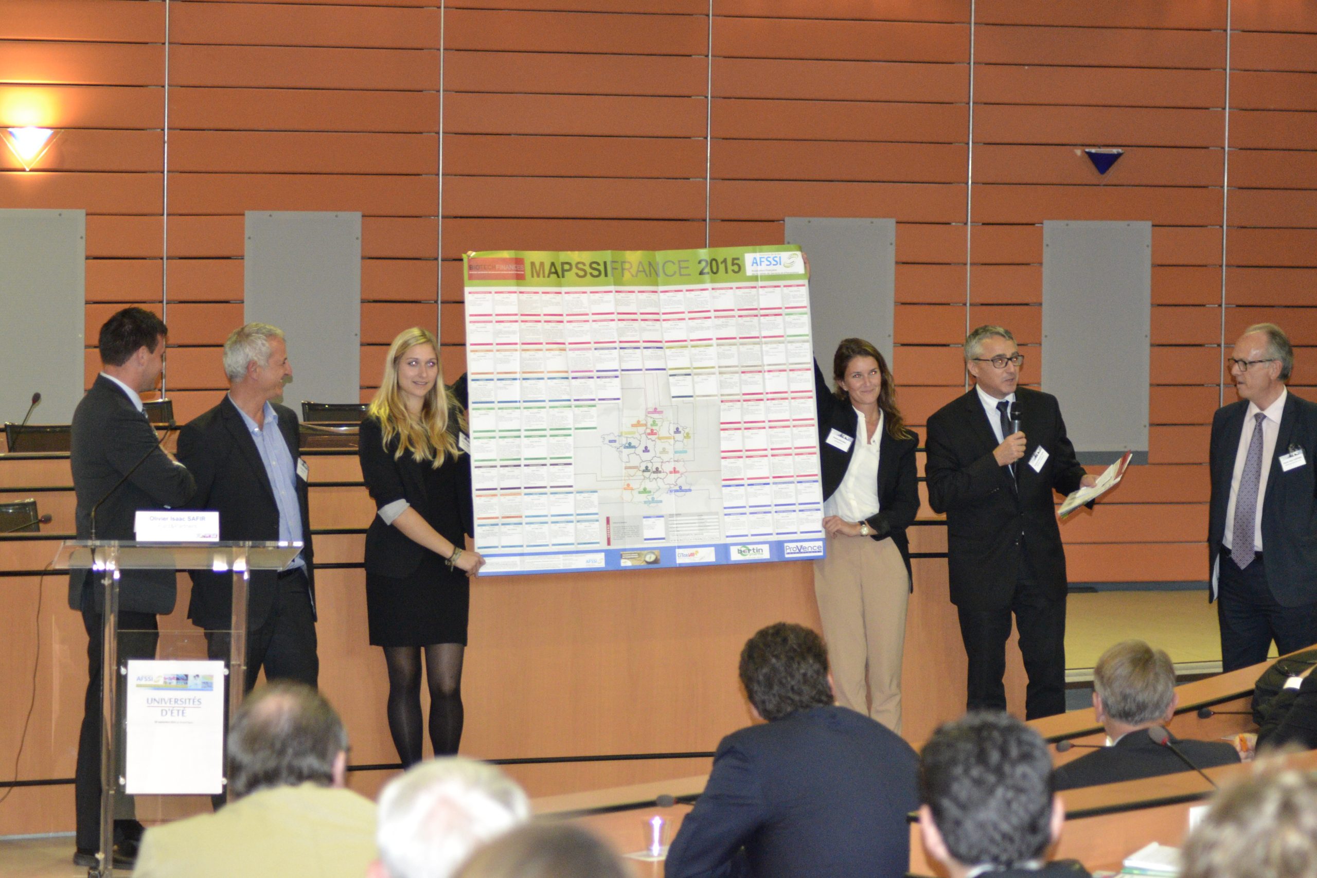
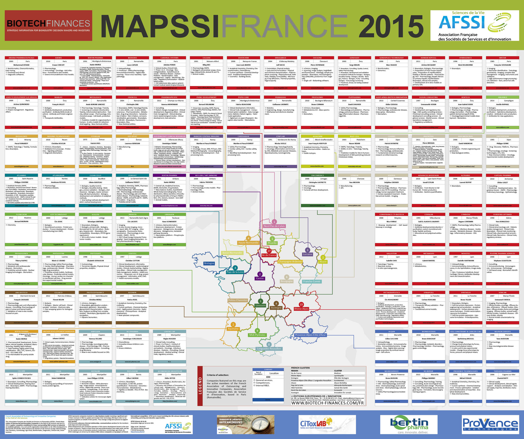
Sponsor MAPSSI

Affichez-vous sur l’édition 2017-1018 de la MAPSSI

Véritable mine d’informations – plébiscitée par tous les acteurs et opérateurs des sciences de la vie – la MAPSSI est un poster de 135cm par 115cm répertoriant les forces vives françaises des sociétés de services et d’innovation en Santé. La MAPSSI est éditée en partenariat avec Biotech Finances.

La MAPSSI sera à imprimée à 3000 exemplaires (soit 0,50€ par personne touchées)

Elle sera distribuée aux 400 participants des journées AFSSI et bénéficiera d’une mise en lumière particulière envers l’écoystème

3000ex

3000 exemplaires imprimés,
soit 0,50€ le contact ciblé

0,50€

Offres d’EXPOSITION

  • Mise à disposition de votre stand sur 2 jours, 1 table, 4 chaises, 1 présentoir, électricité, accès wifi, éclairage, signalétique lettrée
  • Votre logo, lien et présentation sur notre rubrique ‘Exposants‘.
  • Votre logo dans le programme papier de la manifestation (support promotionnel diffusé en amont du jour J et remis sur place aux participants).
  • Mise à disposition de vos plaquettes aux endroits stratégiques de la manifestation.

Sponsor GOLD

Titre

Aller en haut