Posters scientifiques2025-10-03T11:41:37+02:00

Présentez un poster scientifique aux AFSSI Connexions 

Session réservée aux membres AFSSI

Les AFSSI Connexions sont l’occasion de découvrir les innovations proposées par les membres de l’AFSSI !
Les entreprises partenaires de vos projets de R&D

Un jury d’experts

Pierre-Emmanuel GLEIZES

Directeur

Genotoul Genopole Toulouse

Oliver LOGET

Président & CEO

CapEval Pharma

Susanna MALMSTRÖM

Directrice Alliances

Cilcare

Patrick VINCLAIR

VP, BioServices & Welfare Department

EVOTEC

Félicitation à nos lauréats 2025 !

Le prix du jury

Le jury a décerné le prix du meilleur poster à Charles River Laboratories pour son poster intitulé « Innovative collaborative study involving in vitro maturation of oocytes and whole embryo transfer in the mouse », représenté par Marie Bouressam.
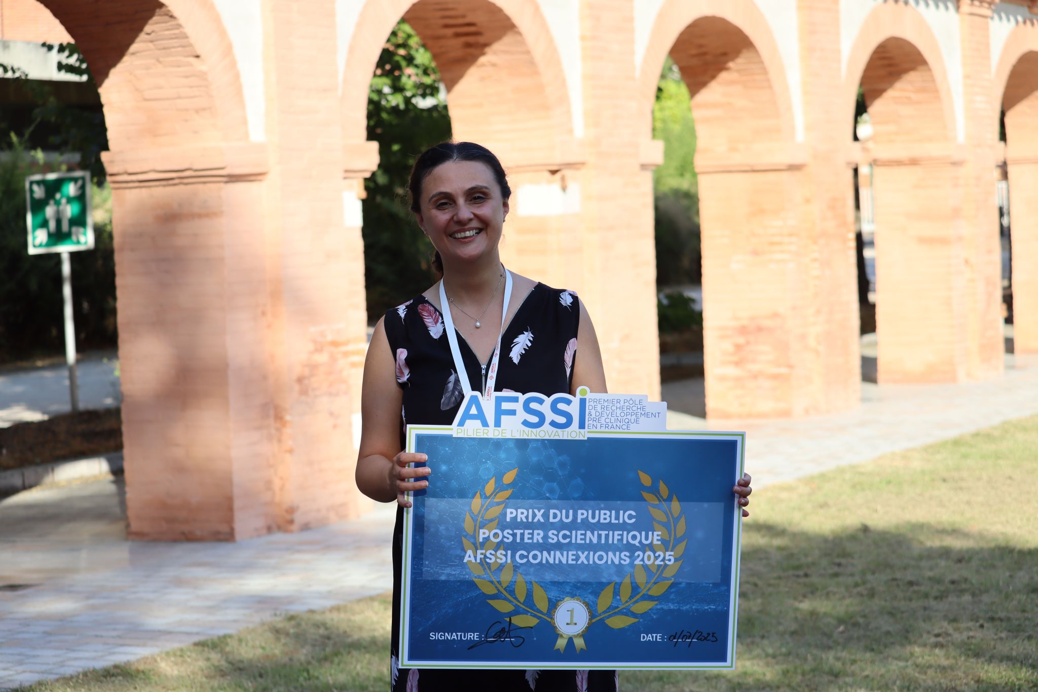
Prix du jury AC2025 : Charles Rivers laboratories

Le prix du public

Le public a récompensé G.CLIPS biotech, représenté par Rosie Dawaliby, PhD et Souad GUESSAS pour son poster « Development of conformational and tissue specific antibody against a GPCR as potential drug candidate in immuno-oncology »

Prix du public AC2025 : G.CLIPS biotech laboratoriesPrix du jury AC2025 : Charles Rivers laboratories

Découvrez les sociétés soumettant un poster scientifique pour l’édition 2025

Probing Nanoscale Viscoelasticity in Living and Biomimetic Systems

Ophélie THOMAS- -CHEMINa, Véronique LACHAIZEa, Arthur GAVEAUa
a : A5 Science, Technopole de l’environnement Arbois-Méditerranée, Aix-en-Provence, France – www.a5-science.com

The mechanical characterisation of cells in regard of their microenvironment is increasingly recognised as a strategic driver of innovation in biomaterials and organoids, mechanosensitive diagnostics and personalised medicine, as these properties regulate critical biological processes, including cell differentiation or migration and tumour invasion¹.

Cells, tissues and biomaterials behave as dynamic viscoelastic systems, whose mechanical properties cannot be reduced to mechanical elasticity². To address this complexity, A5 Science performs dynamic micro-rheology using atomic force microscopy (AFM) coupled with dynamic mechanical analysis (DMA), enabling high-resolution extraction of G′ moduli (accounting for the elastic behaviour of the sample) and G″ moduli (accounting for the viscous behaviour of the sample) over a wide range of frequencies in liquid conditions. This sensitive approach reveals subtle mechanical shifts at the molecular scale, such as the rigidification of polymer networks induced by metal ions³, the impact of point mutations or the denaturation of proteins induced by pH⁴. DMA-AFM is emerging as a powerful tool for studying how environmental signals shape the mechanical landscape of biological systems, with great potential for biomedical research and innovation.

AFM coupled with DMA stands out as a non-destructive, multi-scale and sensitive tool for biomechanical analysis, tissue engineering and molecular biophysics. Using this approach, we are demonstrating its ability to distinguish cancer cells from non-malignant cells based on their early nanomechanical signatures⁵, and to accurately monitor the gelation and stability of hydrogels under a variety of experimental conditions⁶. The comparative analysis of matrices such as PEGDA and Matrigel also illustrates the ability of this technique to reveal composition-dependent mechanical behaviours.
Performed in a controlled liquid environment, A5 Science analysis opens up new prospects for nextgeneration mechanophysical diagnostics.

References:
(1) Cross, S. E.; Jin, Y.-S.; Rao, J.; Gimzewski, J. K. Nanomechanical Analysis of Cells from Cancer Patients. Nat. Nanotechnol. 2007, 2 (12), 780–783. https://doi.org/10.1038/nnano.2007.388.
(2) Gardel, M. L.; Nakamura, F.; Hartwig, J.; Crocker, J. C.; Stossel, T. P.; Weitz, D. A. Stress-Dependent Elasticity of Composite Actin Networks as a Model for Cell Behavior. Phys. Rev. Lett. 2006, 96 (8), 088102. https://doi.org/10.1103/PhysRevLett.96.088102.
(3) Zhu, S.; Wang, Y.; Wang, Z.; Chen, L.; Zhu, F.; Ye, Y.; Zheng, Y.; Yu, W.; Zheng, Q. Metal-Coordinated Dynamics and Viscoelastic Properties of Double-Network Hydrogels. Gels 2023, 9 (2), 145. https://doi.org/10.3390/gels9020145.
(4) Li, X.; van der Gucht, J.; Erni, P.; de Vries, R. Active Microrheology of Protein Condensates Using Colloidal Probe-AFM. J. Colloid Interface Sci. 2023, 632, 357–366. https://doi.org/10.1016/j.jcis.2022.11.071.
(5) Thomas- -Chemin, O.; Séverac, C.; Abidine, Y.; Trevisiol, E.; Dague. AI Classification of Normal and Malignant Cells on the Basis of Their Viscoelastic Properties. Submitted to Biophysical Journal. 2025.
(6) Nalam, P. C.; Gosvami, N. N.; Caporizzo, M. A.; Composto, R. J.; Carpick, R. W. Nano-Rheology of Hydrogels Using Direct Drive Force Modulation Atomic Force Microscopy. Soft Matter 2015, 11 (41), 8165–8178. https://doi.org/10.1039/c5sm01143d


Microalgae-produced antibody as effective as those produced in mammalian cells

Author : Fabien Menissez, Business Development manager

Abstract :

With the rise of chronic diseases such as cancer, autoimmune and infectious diseases, which affect or kill millions of people worldwide, the demand for innovative and specific antibodies as diagnostic tools or biologics has increased significantly.

Currently, these antibodies are produced using mammalian cells as biofactories. However, this type of production has drawbacks such as high production costs, heterogeneous production, instability and potential risk of viral contamination. These high costs limit access to these innovative therapies to only 5% of the world’s population, and to 50% of the population for diagnostics.

Therefore, it is essential to develop alternative cellular biomanufacturing methods that reduce the cost of production of these antibodies while improving their quality and efficacy, thus facilitating patient access to these innovative treatments.

Bioinspired by the ocean, ALGA BIOLOGICS is a Deeptech Greentech company based in Normandie, France developing an innovative platform for antibody production using microalgae as cellular biofactories. Our team has recently demonstrated the ability to produce, on a 200 liter scale, microalgae-based antibodies with a functional profile identical to those produced in mammalian cells.

The company’s latest results on the biological activities and quality of various antibodies produced in microalgae will be presented.

The company is supported in its ambitions by the French government through the France 2030 program under the Acceleration Strategy for Bioproduction and Biotherapies and by the ADD (Aide au Développement Deeptech) of BPI France.

From 3D bioprites models to in-vivo proof of concept : reprogramming of tumor cells into antigen-presenting cells as a new immunotherapy strategy

Abstract : 

The REPRINT project aimed to develop a revolutionary gene therapy based on in vivo reprogramming of immune cells to restore anti-cancer immunity. As part of this Eurostars consortium, Antineo and Asgard Therapeutics have joined forces to provide preclinical proof-of-concept for Trojan-DC, an adenovirus carrying a sequence designed to reprogram cancer cells into antigen-presenting cells.

To achieve this objective, Antineo has developed 3D bioprinted models using human cell lines. This technology enabled to assess the reprogramming rate of cancer cells by Trojan-DC via 3D intra-object injection, and to determine the kinetics of vector reprogramming in the SK-LMS-1 (Leiomyosarcoma) cell line. We then compared these results with the in vivo SK-LMS-1 mouse model : we were able to show that Trojan-DC reprogrammed tumor cells into dentric like-cells and that our 3D bioprinted objects offered predictive results comparable to those obtained in animals.

Toxicological Assessment of NBE’s & NCE’s

Olivier Loget, CapEval Pharma

Abstract :

The primary objective of preclinical studies for new therapeutics or prophylactics is to determine whether they are sufficiently safe to proceed to first-in-human clinical trials and to evaluate their efficacy. These preclinical experiments typically assess toxicity, potency, and immunogenicity. This abstract focuses on Good Laboratory Practice (GLP)-compliant safety (toxicology) assessments for new biological entities (NBEs) and vaccines, and compares them to those for new chemical entities (NCEs).

Such studies are usually performed in selected laboratory animal species. In line with animal welfare principles (the 3Rs: Replacement, Reduction, and Refinement), efforts are being made to minimize animal use. However, the complex mechanism of action (MoA) of vaccines and NBEs often necessitates in vivo testing. While the number of animal studies can be reduced, they remain critical for evaluating safety. We are not yet in a fully “animal-free” research and development era.

Vaccine safety is particularly important, given that they are often administered to infants. The vaccine development process is lengthy—typically involving 1–5 years of initial research, followed by clinical and pharmaceutical development. Overall, licensure may take 15–20 years. Some vaccines, such as for malaria, have required over 30 years of research and dozens of candidates.

In contrast, urgent situations—such as emerging infectious diseases—can accelerate development, as seen during the COVID-19 pandemic. This highlighted the importance of being prepared for rapid development and the need for rigorous safety assessments throughout all phases. The pandemic also underscored the necessity of learning from past experiences to improve scientific methods and global communication strategies.

Innovative collaborative study involving in vitro maturation of oocytes and whole embryo transfer in the mouse

Marie Bouressam, PhD, DABT – Charles River Laboratories, Lyon

Infertility is a disease of the male or female reproductive system, defined by the failure to achieve pregnancy after attempts for 12 months. In Vitro Fertilization (IVF) is part of the assisted reproductive technology and is a complex process involving retrieval of eggs from the ovaries after hormonal stimulation, followed by the fertilization step in vitro.

IVF protocols require ovarian stimulation in order to induce growth and maturation of the oocytes. This hormonal stimulation is associated with risks such as excessive growth of the ovaries, ovary twist and/or ovarian hyperstimulation syndrome. In order to reduce this burden, innovative technologies are being developed to suppress the hormonal stimulation phase.

A new medical device has been developed to allow in vitro maturation of oocytes and therefore suppress the ovarian stimulation phase.

The aim of this study was to induce in vitro maturation of mice oocytes and collect data from pseudopregnant mice reimplanted with embryos produced using the in vitro maturation technique.

First, immature oocytes were collected from B6CBAF1/J mice and cultured in basal medium for approximately 54 hours in the presence of different hormones to induce maturation. Following maturation, oocytes were fertilized using sperm, cultured up to the 2-cell stage and then reimplanted in pseudopregnant CD-1®IGS mice. Reimplanted mice were monitored during gestation and allowed to litter. The dams and pups were submitted to regular physical examinations and necropsied shortly after littering.

This preliminary study confirmed the possibility to obtain viable pups following in vitro maturation of oocytes and fertilization, and subsequent 2-cell stage implantation. This alternative IVF method offers a more natural way of obtaining oocytes without the burden and risks associated with hormonal ovarian stimulation.

Challenges of rat explant culture for a reliable ototoxic ex vivo model for drug screening

Authors : Carolanne COYAT1*, Karine TOUPET1*, Sylvie PUCHEU1*, Gaëlle NAERT1*

  1. Cilcare SAS, 34080 Montpellier France

Cisplatin is one of the most used chemotherapy medications, but its severe ototoxic consequences resulting in irreversible damage to hair cells and hearing impairment, limits its clinical application. The exact mechanism leading to ototoxicity remains unclear. However, cisplatin is well known to induce inflammation, oxidative stress, and apoptosis in the cochlea. This apoptosis takes place in hair cells, principally in the outer hair cells (OHC). In line with 3R principles, reliable in vitro models are of interest and need to be carefully developed. Regarding the cisplatin models, cochlear explant models allow a better selection of therapies against cisplatin’s side effects, allowing drug screening and dose response studies to determine the most efficient drug and its concentration to counteract cisplatin ototoxicity. This ex-vivo model allows to limit animal use, by confirming the therapeutic strategy only with an efficient drug at an efficient dose, that could be used in later stages in vivo.

The objective of this study was to develop the most accurate ototoxic ex vivo model, using rat cochlear explants. In addition, the technical challenges are also presented and discussed.

As a result, we worked with different parameters characterizing the cisplatin explant model: age of the pups, the composition of culture medium, the dissection method, cisplatin concentration, duration of cisplatin exposure and culture period. In addition, to clearly define the effects of cisplatin on cochlear cells, several markers (Myosin 7A, Phalloidin, Hoechst, b-tubulin, V-Glut 3 and Sox2) were used and validated. This allowed the visualization of the various structures of the cochlea, such as hair cells, supporting cells, fibers, and neurons. Depending on the target structures, it is possible to choose more or less severe in vitro models of toxicity induced by cisplatin.

In parallel, a method of image acquisition using a laser scanning confocal microscope as well as histological analysis methods based on a qualitative and quantitative assessment of hair cells (scoring of hair cell organization and counting of hair cell numbers) were developed and validated to provide the most relevant and reliable method to analyze cisplatin effects on the rat cochlea explants.

The development of reliable and consistent rodent explant cultures offers significant advantages for investigating cisplatin’s mechanisms of toxicity and for the development of novel therapies. Preclinical testing is indeed a critical phase in the development of new drugs, making it essential to continually refine and expand tools, including in vivo and in vitro models, to advance the progress of new treatments. In this study, we assessed the toxic effects of cisplatin, but similar investigations can be conducted for various other ototoxic drugs, such as gentamicin, kainate, and more.

Dev4All osteoarthritis Platform — One Question, One Model, One Partner

Authors: Coralie Meurot, Céline Martin, Margot Vieubled, Indira Toillon, Claire Deldycke, Valentine Chéron, Soline Trombetta, William Sibran, Daniel Sanchez Lopez, Revital Rattenbach, Francis Berenbaum

Osteoarthritis (OA) requires therapeutic approaches that address both pain and structural preservation. Dev4All routinely establishes and validates two complementary rat models that map directly onto the key clinical questions in osteoarthritis:

  • MIA model → pain & acute synovitis
  • ACLT/ pMMx model → synovitis & cartilage structure

Objective — Showcase how our question-driven model selection strategy delivers decision-making data for partners developing OA therapeutics.

Methods — A test compound and a corticosteroid (standard of care) were evaluated head-to-head. End-points: pain (von Frey), synovitis (Krenn or Obeidat), cartilage integrity (OARSI).

Results:

  • MIA: Both treatments achieved comparable analgesia with von Frey testing indicating a sustained reduction in mechanical hypersensitivity. Histologically, both treatments reduced inflammation (Krenn inflammation subscore), but the corticosteroid had no impact on synovial cell density and only mild improvement on layer thickness. Only the test compound normalized synovial histology, lowering the total Krenn score from high- to low-grade. A correlation was found between total synovial score and von Frey results (r² = 0.83 pain vs synovitis, p0.001).
  • ACLT/pMMx: The test compound outperformed the corticosteroid on every Obeidat sub-score (hyperplasia, cellularity, and fibrosis) and was the sole agent to confer significant cartilage protection (-40.9 % OARSI score vs vehicle, p<0.01).

Conclusion — Two models, one integrated answer. The corticosteroid reproduces clinical experience—pain relief without structural benefit—whereas the test compound combines analgesic and clear preservation against joint structural changes (synovium and cartilage). By aligning the model with your lead question (pain, inflammation, or cartilage preservation), Dev4All delivers translational, go/no-go data.

Our capabilities at a glance:

  • >30 OA studies completed
  • In-house histopathology & pain behavior scoring
  • Rapid study start-up (≤4 weeks)

DEVELOPMENT OF CONFORMATIONAL AND TISSUE SPECIFIC ANTIBODY AGAINST A GPCR AS POTENTIAL DRUG CANDIDATE IN IMMUNO-ONCOLOGY

Authors: LISA FRELAT¹, FLORIANE AUDEBERT¹, RACHEL LATGER¹, HELICIANE PALENZUELA¹, ROSIE DAWALIBY¹

¹ G. CLIPS Biotech, Labège, France

G protein-coupled receptors and more specifically chemokines receptors are widely implicated in several oncological indications. They can play a role in many processes such as tumor growth, angiogenesis, chemotaxis, and metastasis. Thus, they have great potential as drug targets in immuno-oncology.

Our project is to develop a tissue selective and conformational specific drug candidate against a chemokine receptor implicated in hepatocarcinoma. The GPCR of interest for this study is RD06 (anonymous name). RD06 is highly expressed in the liver and is known to be involved in several cancers, notably liver cancer,
where it has already been considered a key factor in the progression of HCC.

To achieve this goal, the first step was to obtain the antigen of interest (RD06 stabilized in physio pathological conditions) and use it for antibody discovery. G.CLIPS Biotech has developed an enabled technology to produce in vitro specific conformations and native lipid environments on purified membrane proteins. This innovative technology overcomes one of the problems encountered in the development of antibodies targeting membrane proteins. In this study, a combination of different propriety methods was used: (1) Purification of recombinant RD06 with G.mixes® in bicelles and verification of the conformations of the protein with G.Select®; (2) Reconstitution of RD06 in nanodiscs in a specific environment representing liver and lipids raft, and finally ;(3) Pharmacological profiling of benchmark ligand on the receptor in different environment and conformation; (4) Preliminary yeast display experiment on the antigens obtained above for antibody discovery.

Our data show that our technology can unlock selection of conformation/environment sensitive ligand/drug candidates. This finding pave the way towards de-risked candidates on efficiency/ toxicity balance and open opportunities towards development of innovative and selective therapies for unmet medical needs.

Key words: efficiency, toxicity, drug development membrane protein, GPCR, stabilization, conformations, purification, cancer, environment, lipids, nanodiscs, bicelles.

Co-developing a Smart Project and Client Management Platform

In a shared commitment to innovation and operational excellence, Vibiosphen partnered with Gencovery to co-develop an integrated application that enhances project management, client engagement, study planning, and delivery.

Vibiosphen is a French biotechnology company founded in 2014, with over 10 years of expertise in translational research and development. The company specializes in preclinical services and collaborative R&D, operating at the crossroads of human health, animal health, dermocosmetics, and nutraceuticals.

Founded in 2020, Gencovery is a software company that provides a data platform to streamline analysis, integrate artificial intelligence, and accelerate innovation and productivity in life sciences.

Until now, Vibiosphen uses these separate tools with manual intermediates steps:

  • A CRM for client management (quotes, orders, invoicing)
  • An electronic lab notebook (ELN) for study execution (protocols, raw data, reports)
  • Equipment-provider software with Excel software to process internal data

This fragmented ecosystem caused inefficiencies, delays, and made it difficult to manage client relationships and project lifecycles. Vibiosphen believes that more flexible, AI integrated digital solutions are needed to connect tools, streamline operations, and maximize the value of data.

While Gencovery was developing a project management system based on Agile methods, this collaboration enriched the tool with features tailored to the CRO environment and unlocked new co-valorization opportunities.

The jointly developed application offers:

  • End-to-end digitalization of workflows (with CRM integration)
  • Lean visual management with customizable dashboards and Gantt-like tracking
  • Streamlined communication via automated notifications, task coordination, and
    client access to project status
  • Integration of technical study data with business processes

This partnership supports Vibiosphen’s digital transformation while extending Gencovery’s reach in the life sciences sector.

ISCHEMIA-REPERFUSION CELLULAR MODELS TO IDENTIFY MITOCHONDRIA-TARGETED THERAPEUTIC SOLUTIONS

Léa Zennaro, Nelly Buron, Cécile Martel, Mathieu Porceddu, and Annie Borgne-Sanchez
MITOLOGICS SAS, 172 rue de Charonne, 75011 Paris, France

aborgne.sanchez@mitologics.com

Mitochondria play a key role in intracellular energy production and controlling cell life and death. During ischemia, lack of nutrients and oxygen provokes metabolism inhibition and impairment of mitochondrial activity. During reperfusion, the sudden return of oxygen overwhelms mitochondrial capacity decreased by ischemia, leading to free radical production and cell death. As mitochondrial impairment constitutes the principal cause of tissue damage related to ischemia-reperfusion, we investigated the potential of mitochondrial transplantation as therapeutic approach to overcome mitochondrial default.
For this purpose, we set up two in vitro cellular models of kidney and brain ischemia-reperfusion, by inducing oxygen, glucose and glutamine deprivation in kidney LLC-PK1 and brain Neuro-2a cells. At reperfusion (return to normal culture conditions), the cells were directly treated with mitochondria, isolated from mouse liver (Porceddu et al., 2018; Buron et al., 2024). The ability of this strategy to rescue cell metabolism and survival after reperfusion was evaluated by assessing global oxygen consumption and cytotoxicity using our multiparametric assays.

The measures revealed that mitochondria addition significantly enhanced the respiration capacity in cells cultured in both normal and ischemia-reperfusion conditions. The mitochondrial recovery in pathological models was associated with an improvement of cell viability. These results demonstrated that our cellular models of kidney and brain ischemia-reperfusion can be used to identify new therapeutic solutions aiming to restore mitochondrial activity.

Buron N, Porceddu M, Loyant R, Martel C, Allard JA, Fromenty B, and Borgne-Sanchez A. Drug-induced impairment of mitochondrial fatty acid oxidation and steatosis: assessment of causal relationship with 45 pharmaceuticals, Toxicol. Sci., 2024. doi.org/10.1093/toxsci/kfae055.

Porceddu M, Buron N, Rustin P, Fromenty B, and Borgne-Sanchez A. In vitro assessment of mitochondrial toxicity to predict drug-induced liver injury. In book: Methods in Pharmacology and Toxicology – Drug-induced liver toxicity, 2018 Eds. M. Chen & Y. Will, Springer LLC, New York (NY, USA).

TCR-Based Discoveries : a Single-cell Platform for PrecisionT CellTherapy

Barennes, H.P. Pham, M. Touati, V. Quiniou | Parean Biotechnologies

Cytotoxic T cells play a key role in recognizing and eliminating cells presenting abnormal antigens. Biotech and Pharma are looking for new modalities, and leverage TCR as a new tool, in oncology and auto-immune diseases.
However, their very low frequency makes detecting antigen-specific T cells challenging. This low abundance is a major obstacle to the development of effective TCR based therapy. Our platform overcomes this limitation by amplifying and identifying these rare T cells, which often remain undetected by traditional methods.

Using advanced single-cell screening that considers gene expression, cellular pathways, cell expansions and antigen specificity, we can identify these cells and design antigen-specific TCR repertoires.

Our Insilico tools supports the screening and selection of TCR candidates, federating predictive scores for potency, cross-reactivity or immungenicity,
This approach offers new opportunities for the discovery of TCR-based therapy candidates, particularly for neo-antigens, tumor associated antigens, and viral antigens.

Solutions for Overcoming and Leveraging Versatile Efforts Against Microbial Resistance (SOLVE-AMR): a synergistic collaboration for a fully humanized monoclonal antibody (Mab) proposed in a viral infection

Regis Villet (1) , PhD; Patrick Larcier (2), PharmD

(1) BioAster, Lyon, France ; (2) Cencora PharmaLex, Paris, France

A success virtual case study from the S.O.L.V.E. Anti-Microbial Resistance (AMR) Initiative presenting a synergistic collaboration in the development of a biological product in a viral infection from an US-based biotech company.

Their objectives were to:

  1. define the nonclinical and the early phase clinical development plans,
  2. set-up this early clinical study in Europe and
  3. then possibly use the exploratory efficacy data from an early clinical trial for granting the PRIME Designation from the European Medicine Agency (EMA), the EU Orphan Drug (OD) Designation and pursue the development of a fully humanized Mab, in patients suffering from a viral infection.

Based on successful outcomes, such a collaboration highlighted the need for Small- & Mid-Size Enterprise (SME) companies to get support from experienced service providers in the field of AMR.

3D-Living System : Innovative preclinical and physiological models to dissect molecular, cellular and tissular mechanisms in the Central Nervous System.

Dr. Amandine Roux

Research PIECES is a Deeptech startup of Biotechnology.

The goals of the startup are to understand the physiology and the physiopathology of the Nervous System (central and peripherical) through Research, Development & Treatment to fight against neurological diseases (neurodegenerative, cancer, infectious diseases, etc.).

For that, we provide biological and innovative systems to dissect molecular, cellular and tissular mechanisms in a physiological 3D-living tissue environment.
Our physiological models are tailor made, no artificial (versus organoid/ bioprinting), dynamic (versus in vivo) and integrated (versus in vitro), with the advantages of in vivo and in vitro without the disadvantages.

By providing robust and translatable systems close to human physiology we allow avoiding therapeutical molecules fail in clinical trial phases.
As also, an alternative to animal models, by following the 1st « R » of the 3R European rule, our 3D-Living System also permit to drastically decrease the number of animals used in Research.

They can be used for the different steps of the early drug discovery stages (screening, lead of molecules, target validation, etc.), tests of toxicity and in many therapeutic areas, as neurodegenerative diseases (Parkinson, Dementia with Lewy Bodies, etc.) or cancer.
They can also permit to study the impact of the environment on the CNS by studying the role of pesticides, endocrine disruptors, or neurotoxins, but also the Biophysical parameters (frequency, radiation, etc.).
Our products/services are designed for all the researchers from Academia (CNRS, Inserm, etc.) to Pharmaceutical Industry (CRO, CDMO, etc.), and Army
(as antidote to neurotoxins), animal health (veterinary products) and cosmetics. Research PIECES has the innovation to find the future therapeutic molecules of tomorrow to permit a better knowledge of CNS to cure and prevent neurological diseases

Advancing Alzheimer’s disease models for preclinical drug testing

Authors: Allouche A.1, Tallandier V.1, Kridi S.1, Dier L.1, Colin J.1, Robelet Sandra, Violle N.1
ETAP-Lab, 54500, Vandœuvre-lès-Nancy, France

Alzheimer’s disease (AD) is the most common cause of dementia, complete disease-modifying treatments are yet to be fully attained. The development and commercialization of new drugs is slow and expensive process and current approaches targeting the too-late stages of AD have had no consistent clinical benefit. Indeed, development of predictive and robust preclinical models, recapitulating early neuropathological features of AD are of urgent need. Here, we report the progress in our development of high throughput in vitro models that captures AD characteristics using physiopathologically relevant culture conditions.

Assessment of cellular phenotypic changes (neurons or glial cells from rodent or human induced pluripotent stem cells (iPSCs) induced after an exposure of human amyloid oligomers (AβO and TauO) were performed using 2D-cultures, brain-on-chip (BoC) models, and a high content imaging system.
These oligomeric preparations were prepared in-house from human Aβ1-42 and Tau (2N4R) monomers and were well-characterized by various methods (SDS page, dot-blot, 8-Anilino-1-NaphthaleneSulfonic acid (ANS) and Sedimentation Velocity Analytical Ultracentrifugation (SV-AUC) assays).

Our data showed that increasing concentrations of oligomers have been associated with significantly loss of synaptic markers, reduction of neurite outgrowth and ultimately neuron death. They also triggered neuroinflammatory processes via activation of astrocytes. Induced neurodegeneration and neuroinflammation were successfully attenuated by pharmacologic compounds, including monoclonal antibodies, which have produced encouraging therapeutic results in clinical trials.

In conclusion, we characterized many experimental situations to better understand the mechanisms underlying AD, and targeting oligomers or their harmful effects which should give rise to new therapeutic interventions. Complexifying of in vitro models, combining neuron and glia, using BoC devices and targeting both amyloid and tau for development next generation AD prevention could be of great interest.

Integrated High-Throughput Protein Expression Platform combining cell-free and bioluminescence technologies with Artificial Intelligence (AI)

Authors : Y. Abdelaziz, M. Gransagne, K. Jondot, J. Spiaczka, B. Tillier

Abstract:

With the growing use of artificial intelligence (AI) to optimize protein expression, particularly in cell-free systems, new challenges have arisen in efficiently generating high-quality, protein-specific data. Traditional methods often require multiple rounds of experimentation, making them both time-consuming and costly. To address these limitations, we are developing an AI-driven Design of Experiments (DOE) framework. Our approach leverages protein sequence data to extract key parameters, which are then processed by a recommendation system to propose optimal experimental conditions. This enables faster iterations, greater flexibility, and significant cost and time savings compared to traditional active learning strategies.

By combining this approach with our next-generation bioluminescence technology, we can rapidly establish optimal expression conditions in our cell-free system for almost any type of protein. Once these conditions are established, our platform has the capability to express hundreds to thousands of DNA sequences, providing purified protein samples ready for initial characterization in less than 48 hours.

Here, we present a case study demonstrating a significant improvement in the expression yield of VHH candidates using our AI-guided cell-free expression system. By optimizing the experimental conditions, such as temperature, buffer composition, and transcription/translation rates, we were able to increase the yield of functional VHHs. This result highlights the power of our integrated platform in rapidly identifying the most efficient conditions for protein production, without the need for time-consuming trial-and-error experiments.

Building on this success, our platform offers a robust and scalable solution for the expression and screening of diverse protein libraries. Our process not only saves valuable time and resources, but also provides a more flexible and reproducible method for selecting protein-based therapeutics, diagnostics, or research tools.

Découvrez les présentations des plateformes dans le cadre du partenariat avec le PUI de Toulouse

Genotoul : un écosystème technologique d’excellence pour les partenariats de recherche et développement en sciences du vivant

Abstract : 

Genotoul représente l’infrastructure de coordination des plateformes technologiques en sciences du vivant du pôle scientifique toulousain. Créé en 1999 et structuré depuis 2009 en groupement d’intérêt scientifique (GIS), il réunit organismes de recherche, établissements d’enseignement supérieur, CHU de Toulouse, collectivités territoriales et pôles de compétitivité, offrant un terrain propice au développement de partenariats entre recherche académique et innovation industrielle.

Genotoul fédère 20 laboratoires plus de 60 plateaux techniques du site toulousain au sein d’un réseau de 14 plateformes spécialisées offrant des expertises et des services de haut:

  • Technologies « omics » et analyses moléculaires : génomique (GET, CNRGV), protéomique (Proteotoul), métabolomique (Metatoul) pour l’identification et l’analyse des molécules à très haut débit.
  • Innovation thérapeutique : recherche et optimisation de molécules d’intérêt thérapeutique ou biotechnologique (PICT) par criblage moléculaire, identification de biomarqueurs (WeMet).
  • Imagerie et modélisation : visualisation du vivant de la molécule à l’organisme (TRI), caractérisation de modèles animaux, végétaux et organoïdes (ANEXPLO, Phenotoul, TOP).
  • Support scientifique : bioinformatique et biostatistiques (Bioinfo, Biostat), biobanques (CRBh, CNRGV), accompagnement éthique et réglementaire (Societal).

Les règles de fonctionnement de ces plateformes sont reconnues par la labellisation IBiSA au plan national. Elles sont certifiées par des normes qualité (ISO 9001:2015, NFX50-900) et fonctionnent sur la base d’une tarification clairement établie pour les acteurs académiques et privés.

Ainsi, forte de 25 ans d’expérience, Genotoul offre aux entreprises un accès privilégié aux technologies de pointe du monde académique pour développer des bioprocédés, des approches de médecine personnalisée, identifier de nouveaux biomarqueurs et accélérer l’innovation en biotechnologie et santé humaine.

Technology and expertise platform of the Institut de Chimie de Toulouse (ICT)

Characterization of nanomaterials by Field-Flow Fractionation techniques (FFF): An example of polyoxazoline nanovectors analysis for photodynamic therapy

Abstract : 

FFF (Field-Flow Fractionation) techniques fractionate structures between 1nm and a few micrometers. These techniques are close to liquid chromatography but the separative tool is a channel without a stationary phase. It avoid shear forces and preserve native structure. FFF combines the separating effect of laminar flow going through the channel and a perpendicular force to this flow. The type of separation (size, density, electrical charge, chemical function) depends on the perpendicular force type (eluent flow, centrifugal force, electric force, temperature gradient). Then populations are eluted in online detectors (UV, RI, MALS, QELS) to obtain molar mass, quantification, size and conformation. The most common technique is the Asymmetrical Flow Field-Flow Fractionation (AF4), it fractionates population by hydrodynamic radius.

FFF techniques can be used for medicine to analyze nano-objects. A study of AF4 nano-vectors analysis is presented here: The study use copolymers based on poly(2-alkyl-2-oxazolin) with a coumarin chain end combined with polymeric crosslinking agents, and pheophorbide has been encapsulated as a photosensitizer. This copolymers have been characterized by AF4-MALS-QELS-dRi, Transmission Electronic Microscopy (TEM) and cryo-TEM. This techniques allowed to discriminate the right candidates for the encapsulation of pheophorbide.

(Re)découvrez les lauréats 2024

Le prix du jury

  • La possibilité de présenter son poster lors de la soirée networking
  • Une mise en visibilité sur nos réseaux sociaux
  • Une place offerte au sein du Village AFSSI pour la prochaine édition des AFSSI Connexions
CILcare, lauréat du prix du jury aux AFSSI Connexions 2024

Le prix du public

  • La possibilité de présenter son poster lors de la soirée networking
  • Une mise en visibilité sur nos réseaux sociaux
Atlantic Bone Screen et Enterosys, lauréats du prix du public aux AFSSI Connexions 2024

CILcare, lauréat du prix du jury

Sensorineural hearing loss as a complication of type-2 Diabetes mellitus: evidence of several cellular and neural impairments.

CILcare, lauréat du prix du jury aux AFSSI Connexions 2024

En gagnant le prix du jury, CILcare remporte :

  • Un accès membre Village AFSSI d’une valeur de 690€HT pour la prochaine édition
  • Une prise de parole lors de la soirée networking
  • Une mise en visibilité sur les canaux AFSSI

Abstract

Carolanne COYAT1*, Karine TOUPET1*, Gaëlle NAERT1*, Sylvie PUCHEU1* and Mathieu SCHUE1*.

  1. Cilcare SAS, 34080 Montpellier France

While retinopathy, nephropathy, and peripheral neuropathy are well-established complications of type 2 diabetes, sensorineural hearing loss is increasingly recognized as a comorbidity of this metabolic disease. Several causes have been suggested, including auditory peripheral neuropathy (synaptopathy), which can lead to early-onset hearing loss. No extensive research has been conducted on preclinical models of type 2 diabetes. Our study aims to better characterize the development of SNHL concomitantly with diabetes biomarkers in a diabetic mouse model.

Genetically modified mice with mutations in the leptin receptor gene (BKS(D)-Leprdb/JOrlRj)2 were monitored from 5 to 13 weeks of age to assess the parameters of their diabetes (blood glucose, glycosylated hemoglobin, biochemical analyses) and their hearing, using auditory evoked potentials and terminal histological analyses of the cochlea.

In this model, Leprdb/Leprdb mice exhibited a phenotype of obesity and hyperglycemia, with fasting blood glucose levels of ~4 g/L compared to 2 g/L in heterozygous control mice. Diabetic mice displayed early-onset hearing loss characterized by a significant increase of auditory brainstem response (ABR) thresholds and a significant decrease of distortion product otoacoustic emission (DPOAE) amplitudes compared to the Leprdb/+ control mice. These data correlated with morphological and histological changes in the cochlea.

Here, we present data from a murine model illustrating the detrimental consequences of type 2 diabetes on hearing. This translational preclinical model may be useful for evaluating the efficacy of drug candidates for the preservation and restoration of hearing in type 2 diabetic patients.

Atlantic Bone Screen et Enterosys, lauréats du prix du public

Décrypter l’Axe Intestin-Articulation : Vers de Nouvelles Stratégies Thérapeutiques ou Préventive pour l’Obésité

Atlantic Bone Screen et Enterosys, lauréats du prix du public aux AFSSI Connexions 2024

En gagnant le prix du public, Atlantic Bone Screen et Enterosys remportent :

  • Une prise de parole lors de la soirée networking
  • Une mise en visibilité sur les canaux AFSSI

Abstract

Dans le contexte de la prévalence croissante de l’obésité et de ses comorbidités, la compréhension des interactions entre l’intestin et les articulations devient cruciale. Enterosys, spécialiste de la communication entre l’intestin et les organes périphériques, en collaboration avec Atlantic Bone Screen, expert en recherche préclinique dans les pathologies osseuses et articulaires, propose une offre de service intégrée pour étudier l’axe intestin-articulation. Cette approche innovante vise à explorer le potentiel thérapeutique de nouveaux actifs dans le traitement des pathologies ostéo-articulaires associées à l’obésité.

Nous avons utilisé un modèle murin, des souris C57BL6J mâles de 9 semaines, soumises à un régime hyperlipidique à 60% pour induire l’obésité. L’étude s’est focalisée sur l’évaluation de divers paramètres : métaboliques (gain de poids, test oral de tolérance au glucose, profils lipidiques plasmatiques, poids des tissus adipeux), intestinaux (marqueurs de la fonction de barrière et perméabilité, analyses histologiques) et articulaires (analyses morphologiques par microtomographie (µCT) et histopathologiques).

Les résultats montrent des modifications significatives des paramètres métaboliques et des altérations des marqueurs de la fonction de barrière intestinale.  Des signes d’inflammation et d’atteintes au niveau ostéoarticulaire peuvent être aussi observés, soulignant l’impact de l’obésité sur la santé ostéo-articulaire.

Cette étude met en évidence le potentiel de l’axe intestin-articulation comme cible thérapeutique dans les pathologies métaboliques et dégénératives. Notre offre de service conjointe permettra d’offrir une solution complète pour l’évaluation de l’efficacité d’actifs dans un cadre préclinique, promouvant une approche holistique dans la gestion de l’obésité et ses comorbidités articulaires.

Offres d’EXPOSITION

Sponsor GOLD

Titre

Aller en haut